Chapter 11 · Section III — Screening & Early Detection
Mammography, Ultrasound & MRI
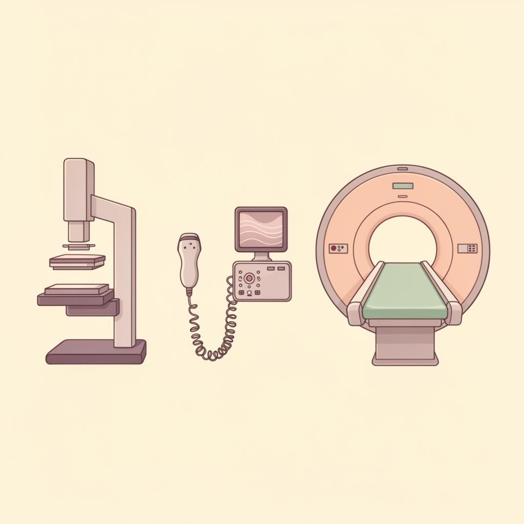
Three different ways of seeing the same tissue. None is the whole truth — together, they offer a fuller picture.
Three lenses on the same tissue — each answering a different question.
Where thermography listens to function, the structural imaging tools — mammography, ultrasound, and MRI — look at form. Each uses a different physical principle (X-rays, sound waves, magnetic fields) to make breast tissue visible. None is universally "best." The right tool depends on your age, your breast density, your risk profile, and the question being asked.
Understanding what each test sees — and what it cannot see — is how you become an informed partner in your own screening, instead of a passive recipient of whichever test is offered first.
Mammography
The X-ray view
Best for
Women 40+ with average-density breast tissue.
Detecting microcalcifications — tiny calcium deposits that can be an early sign of DCIS.
Establishing a structural baseline alongside other tools.
Limitations
Less sensitive in dense breast tissue (Category C & D).
Can miss tumors hidden behind dense glandular tissue.
Ultrasound
The sound-wave view
Best for
Distinguishing solid masses from fluid-filled cysts.
Imaging dense breast tissue where mammography is limited.
Pregnancy, breastfeeding, and women under 40 — no radiation.
Guiding biopsies in real time.
Limitations
Operator-dependent — quality varies with the technician's skill.
Not ideal as a sole screening tool; usually paired with mammography.
Can produce false positives, leading to extra biopsies.
MRI
The contrast-enhanced view
Best for
High-risk women (BRCA1/BRCA2, strong family history, prior chest radiation).
Evaluating the extent of a known cancer before treatment.
Imaging breast implants for rupture or leakage.
Dense tissue when mammography is inconclusive.
Limitations
Highest cost; often requires insurance pre-authorization.
Uses gadolinium contrast — not ideal for kidney concerns or pregnancy.
High sensitivity means more false positives and follow-up scans.
Choosing the right tool
There is no one-size-fits-all screening protocol. The table below offers a starting point — a conversation to bring to your practitioner, not a prescription.
Your situation
Often suggested
Average risk, age 40+, average density
Mammography (annual or every 2 yrs) ± Thermography
Dense breast tissue (Category C/D)
Mammography + Ultrasound, or MRI if very dense
Under 40, no symptoms
Thermography for baseline; Ultrasound if a lump is felt
BRCA+ or strong family history
Annual MRI alternating with Mammography every 6 months
Pregnant or breastfeeding
Ultrasound (and Thermography) — no radiation
Implants present
MRI for implant integrity; Mammography with implant displacement views
Questions worth asking
Before any scan, give yourself permission to be curious. These questions invite the kind of conversation that turns a screening into shared decision-making.
1
What is my breast density category, and how does it affect what this scan can see?
2
What are the benefits, risks, and limitations of this specific test for my body?
3
What happens if something is found — what's the next step, and how long will I wait?
4
Are there non-radiation options that could complement this scan?
5
How will the results be communicated to me, and by whom?
A reflection
"The best screening plan is the one you understand, agree with, and actually keep."