Chapter 10 · Section III — Screening & Early Detection
Thermography Explained
A gentle, radiation-free way to listen to what your breast tissue is doing — long before there is anything to see.
Thermography — sometimes called digital infrared thermal imaging (DITI) — is a screening tool that measures the heat radiating from the surface of your skin. Because heat is a byproduct of metabolism, the patterns it reveals are a kind of map: of circulation, of inflammation, of how your breast tissue is behaving in this season of your life.
It is not a replacement for mammography. It is a different kind of question. Where structural imaging asks "is there a lump?", thermography asks "how is this tissue functioning?" Together, they offer a far fuller picture than either alone.
Four Guiding Principles
Heat tells a story
Every metabolic process — circulation, inflammation, hormonal activity — releases heat. Thermography simply listens to that heat with a high-sensitivity infrared camera.
Function before structure
Where mammography looks for a mass that already exists, thermography looks at how the tissue is behaving. Functional change often precedes structural change.
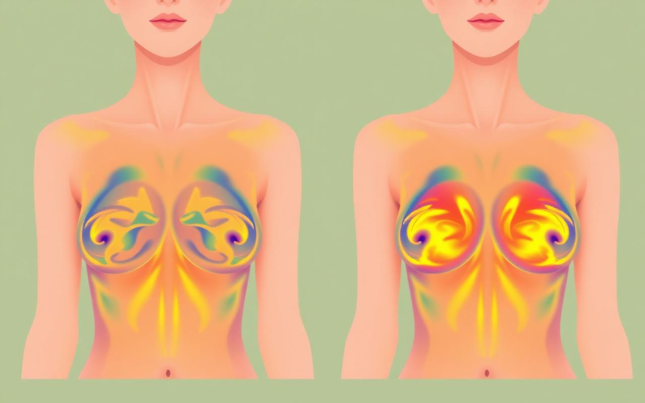
Symmetry is the language
Healthy breasts are thermally symmetrical. Asymmetric heat, vascular patterns, or hot spots can hint at congestion, inflammation, or hormonal imbalance worth exploring.
No radiation, no compression
Thermography is non-invasive. There is no contact, no compression, and no ionizing radiation — making it safe to repeat as often as needed.
Especially helpful for
Women of all ages, including those under 40 when mammography is less informative.
Dense breast tissue, where structural imaging can be limited.
Women with implants, fibrocystic tissue, or a history of biopsies.
Tracking the effects of lifestyle, hormonal, or detoxification work over time.
Pregnant or breastfeeding women — no radiation, no contact.
What it is not
Diagnosing cancer. Thermography does not name disease — it flags physiology that may need follow-up.
Replacing mammography, ultrasound, or MRI. It complements them.
Producing an instant answer. Patterns are most meaningful when compared over time.
What a thermography visit looks like
- 01
Before
Avoid heavy exercise, sun exposure, hot showers, and lotions for several hours before your scan. Wear loose clothing.
- 02
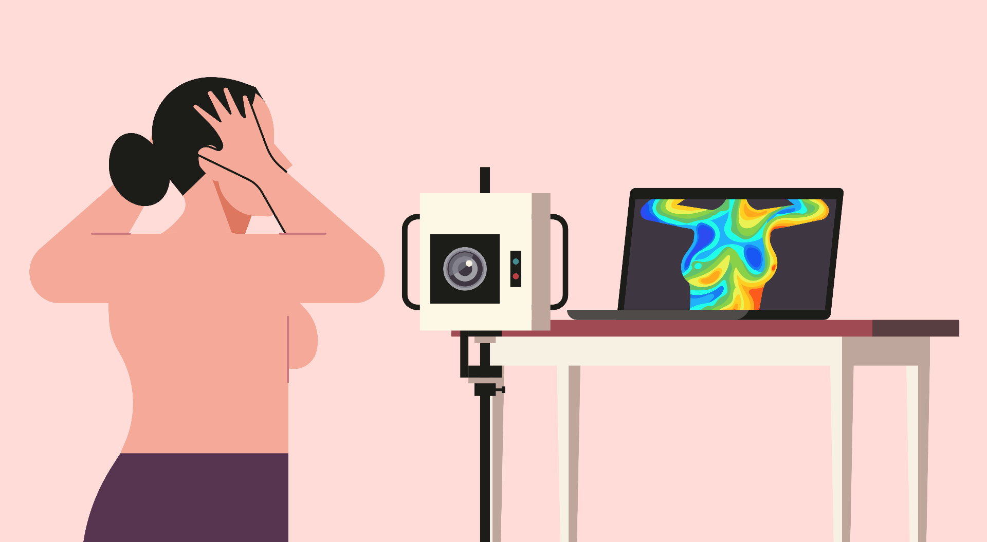
Acclimation
You'll undress from the waist up in a cool, climate-controlled room for about 15 minutes so your skin reaches a stable baseline.
- 03
Imaging
A series of contact-free infrared images are captured from the front and sides. The session is quiet, calm, and takes only minutes.
- 04
Interpretation
Images are read by a board-certified thermologist. You receive a written report with thermal grade, observations, and follow-up timing.
- 05
Baseline & follow-up
A first scan establishes your baseline. A follow-up at three months confirms the pattern is stable. After that, annual scans monitor for change.
A reflection
"Thermography doesn't tell you what is wrong. It tells you where your body is asking for attention — and gives you time to answer."